ss_blog_claim=b2279e430ccb7170410db0a73c6dc7ad

2009/12/17

OF D'S AND F'S

Many people wonder what these acronyms mean when they visit the doctors' clinic...aside from MD there are D's and F's i.e. DPBU, DPBS, DPOGS, DPCP, DPPS, DPBA and FPUA, FPCS, FPOGS, FPCP, FPPS,  etc...

Now, what does the D mean? It stands for DIPLOMATE (DPBU- Diplomate, Philippine Board of Urology).  It is defined as an individual who has earned a diploma or certificate, especially a physician who has been certified by a specialty board (Mosby's Medical Dictionary, 8th edition. © 2009, Elsevier.).  A person with a degree of higher education, a diplomate 
Graduate education A physician who is board-certified in a particular specialty and holds a diploma from a specialty board. (McGraw-Hill Concise Dictionary of Modern Medicine. © 2002 by The McGraw-Hill Companies, Inc.).

Then we come to the F.  It stands for FELLOW (FPUA - Fellow, Philippine Urological Association).   
1) A physician who has attained specified credentials required for admittance to a professional organization.
2) A physician who enters a training program in a medical specialty after completing residency, usually in a hospital or academic setting.
(The American Heritage® Medical Dictionary Copyright © 2007, 2004 by Houghton Mifflin Company).

Here in the Philippines, each medical specialty has an accreditation body that is in charge of evaluating the training institutions as well as its trainees and graduates.  Each training institution is evaluated based on the number of cases, beds, trainees (residents/fellows), equipment and instruments, educational material etc.  For the residents, there is an annual evaluating exam also known as the residency in service exam.

To become a DIPLOMATE, one must be a graduate of an accredited training program before qualifying to take the  diplomate exams.  The type of exams as well as the time as to the eligilibility to take the exam varies on the specialty board.  There are specialty boards that give written and oral exams while there are some that give written, oral and practical exams.  After complying with the requirements and passing these exams, one is a certified diplomate and will be inducted by the specialty board.

To become a FELLOW, one must be a diplomate and then has to apply to the organization where the specialty belongs.  Each specialty organization has their criteria for acceptance and the applicant has to submit the necessary credentials.  Once the criteria is met and credentials have been reviewed and accepted, the applicant will be notified and will be inducted. 


Oath taking led by Dr Nelson Patron, Chairman, Philippine Board of Urology at EDSA, Shangrila during the Philippine Urological Association Annual Convention, Nov 26, 2009
Members of the PBU (L-R) - Dr Ariel Zerrudo, Dr Jesus Benjamin Mendoza, Dr Eduardo Gatchalian


Oath taking


My diplomate certificate from the Philippine Board of Urology

2009/10/06

Research Papers Done

Specialty training is not only full of eight or twenty four hour duties, ward calls, ER calls, operating room procedures, OPD consultations, there is also room for research work.  All training programs whether you are in the medical or surgical specialty, have a required number of research papers to be completed.


Above: Presenting my case report on "Bilateral Ureteropelvic Junction Stenosis in the Upper Moieties of a Complete Duplex Collecting System" during the St Lukes Medical Center Urology Week 2005.  This case report was published in the Philippine Journal of Urology Dec 2005.
Below: All smiles as I won 2nd place during this case report contest.


This was taken during 2005 Philippine Urological Association Annual Convention when I presented a poster on my research paper "Complications of Transrectal Ultrasound Biopsy of the Prostate in the National Kidney and Transplant Institute". 


Letter from the Asia Pacific Association of Pediatric Urologists for the 9th Annual Meeting in Shanghai, China held last December 2007.
I had an oral presention on the paper "Endoscopic Correction of Vesicoureteral Reflux Using Dextranomerhyaluronic Acid Copolymer (Deflux) in the Philippines".


My paper published in an international journal, Journal of Pediatric Urology, Feb 2008.
This is the first reported case in local and international literature.

2009/09/15

VARICOCELE

Varicocele is a clump of enlarged and engorged veins that develops in the spermatic cord within the scrotal sac. The spermatic cord is made up of veins, arteries, lymphatic vessels, nerves, and vas deferens.


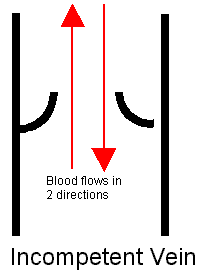
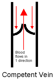
If the valves that regulate bloodflow from these veins are defective (usually from birth), blood does not drain from the testicles efficiently, causing swelling in the veins above and behind the testicles.The pooling of blood in the engorged vessels results in warming of the testis.


A varicocele can develop in one or both testicles, but in about 85% of cases, the left testicle is involved.
A varicocele is found in approximately 15% of  men.  In men evaluated for infertility , varicoceles are found in approximately 40% of patients.

Varicocele affects semen quality because of increased heat to the testicle. The dilated veins allow warm blood from the abdominal cavity to flow around the testicle. This causes overheating of the testicle which then impairs its function. Commonly, a low sperm count, poor movement, and abnormally shaped sperm are found in men with varicoceles. A varicocele surrounding 1 testicle may affect the testicle on the opposite side of the body. A varicocele may also lead to impaired growth of the testicle and thus the testicle on the side of a varicocele may be smaller than the other..

Signs and symptoms

Most men with varicocoeles have no symptoms and they are diagnosed on routine physical examination or during infertility work up.

Signs and symptoms include the following:
  • Pain in the testicle
  • Feeling of heaviness or discomfort in the testicle(s) 
  • Infertility
  • Shrinkage of the testicle(s)
  • Visible enlarged vein or vein that is able to be felt

 Large varicoceles are easily identified on physical examination; they have the classic "bag of worms" appearance surrounding the testis.

Grading of varicocoeles:
  • Grade 1-  varicocele that is palpable only during straining (Valsalva maneuver).
  • Grade 2 - varicocele in which the lesion is palpable without a Valsalva maneuver.
  • Grade 3 - varicocele that is visually detectable
Diagnostics:

Ultrasound is the examination of choice for investigating varicoceles, and it remains the most practical and most accurate noninvasive technique.

Treatment

If the varicocele causes pain or decreased size of the testis (rare) or if the condition is causing infertility (most common), surgery is recommended.

Urologists correct varicoceles by performing a surgical procedure called varicocelectomy.


The Male Infertility Best Practice Policy Committee of the American Urological Association recommends that varicocele treatment should be offered to the male partner of a couple attempting to conceive when all of the following are present


  • A varicocele can be felt..
  • The couple has documented infertility.
  • The female has normal fertility or potentially correctable infertility.
  • The male partner has one or more abnormal semen parameters or sperm function test results.

In addition, adult men whose varicocele can be felt and semen analyses show abnormal findings but are not currently attempting to conceive should also be offered varicocele repair.

A varicocele is the most correctable factor in a male with poor semen quality; therefore, varicocele repair should be considered a viable option for individuals and couples with otherwise unexplained infertility because varicocele repair has been shown to improve semen parameters in most men and possibly improve fertility.

2009/09/13

VESICOURETERAL REFLUX

What is vesicoureteral reflux? (VUR)

The normal urination process involves bladder contraction and urine flow out of the body through the urethra.


With vesicoureteral reflux (VUR), some urine goes back up into the ureters and possibly up to the kidneys. This process exposes the kidneys to infection.


In children, especially those in the first 6 years of life, urinary infection can cause kidney damage. The injury caused by infection to the kidney may result in scars in the kidney and loss of future growth potential or widespread scarring and loss of the normal kidney tissues. Even a small area of scarring in one kidney may be a cause of high blood pressure later in life. Untreated, severe reflux on both sides can result in kidney failure requiring dialysis or kidney transplantation.

VUR is most commonly diagnosed in infancy and childhood after the patient has a urinary tract infection (UTI). About one-third of children with a UTI are found to have VUR.

VUR can lead to infection because urine that remains in the  urinary tract provides a place for bacteria to grow. But sometimes the infection itself is the cause of VUR.

There are undetermined genetic risk factors which may affect the development of VUR. About 34% of patients who have the condition have siblings who are also affected.

Types of VUR:

Primary reflux

The most common cause for primary reflux in children is an abnormality in the section of the ureter that enters the bladder ( intravesical ureter). The intravesical ureter may not be long enough to enable the ureter to sufficiently act as a valve to prevent urine reflux, or the ureter may be inserted abnormally into the bladder.

Other causes of primary reflux include abnormalities in muscle of the bladder, abnormalities in the location of the urethral opening and abnormalities in the shape of the urethral opening. 

Secondary reflux

Secondary reflux is often caused by urinary tract infection which causes inflammation and swelling of the ureter. UTI may cause vesicoureteral reflux or vesicoureteral reflux may promote the growth of bacteria in the urinary tract, causing UTI.

Secondary reflux may also be caused by urinary tract abnormalities like narrowing of the ureter; duplicated ureters; ureterocele) and obstructions from stones or tumors. 


Evaluation

An ultrasound of the kidneys and bladder is done to 1) evaluate hydroneprosis and kidney growth and 2) detect abnormalities that cause reflux.


A renal scan is done to evaluate kidney growth and detect presence of scarring.


A voiding cystourethrogram (VCUG) is performed to determine if an abnormality in the urinary tract is causing reflux. A contrast dye is instilled into the bladder through a catheter and a series of x-rays are taken.


Grading of VUR


Grade I (least severe) to Grade V (most severe):

  • Grade I results in urine reflux into the ureter only.
  • Grade II results in urine reflux into the ureter and the renal pelvis, without swelling of the top of the ureter (hydronephrosis).
  • Grade III results in reflux into the ureter and the renal pelvis, causing mild hydronephrosis.
  • Grade IV results in moderate hydronephrosis.
  • Grade V results in severe hydronephrosis and twisting of the ureter

Mild-to-moderate degrees of reflux (grades I to III) have a good chance of spontaneous resolution with age in over 80% of children. This typically occurs over the span of few years. Unfortunately, we do not know exactly when the reflux will go away for a particular child.
The chance of spontaneous resolution of high grade reflux (IV to V) is much lower.


Treatment

1) Antibiotic prophylaxis for VUR Gr II-III
2) Endoscopic treatment for VUR Gr I-IV
3) Open surgery  for VUR Gr IV-V


Follow-Up

All patients with a history of reflux will have life long monitoring. Even if the reflux resolves, there is still risk of developing kidney insufficiency, hypertension, and pregnancy-related problems.This usually involves periodic visits to the pediatrician and have the following taken: height and weight, blood pressure, and urine analysis. Kidney function can be evaluated by blood tests (creatinine and BUN) or by creatinine clearance or glomerular filtration rate. Occasional ultrasound tests will ensure that kidney growth is on target for age. Female patients should be carefully monitored during their pregnancy.

2009/02/18

UNDESCENDED TESTIS

Undescended testicle, also called cryptorchidism, is a common condition in which one of the testicles is not located within the scrotum. During the eighth month of the mother's pregnancy, the baby's testes migrate from the abdomen, through the groin, and into the pouch that contains the testes (scrotum).

An undescended testicle may be located in the abdominal cavity, in the passageway in the groin (inguinal canal), or in an ectopic location (e.g., superficial pouch in the groin, perineum, upper thigh). This condition is usually present at birth (congenital) and is associated with sterility and an increased risk for testicular cancer if not corrected.

Undescended testicle may occur in approximately 30% of premature males and 3% of full term male infants. In 80% of cases, the undescended testicle migrates into the correct position without intervention during the first year. The condition may involve both testicles in about 10% of cases.

The cause of undescended testicle is not known. If the father or brother ad the conditionm, there is an increased risk. Other risk factors include the following:

* Low birth weight (less than 2500 g)
* Maternal exposure to estrogen during the first trimester
* Multiple birth (e.g., twin, triplet)
* Premature birth (before 37 weeks gestation)
* Small size for gestational age

Diagnosis of this condition is made through physical examination at birth to locate the testis. If one testicle is undescended, the scrotum appears unbalanced. If the undescended testis is felt (palpable) it may not have descended fully, may have descended into a location other than the scrotum (ectopic), or may move in and out of the scrotum through muscle contraction (retractile).

If the testis is non palpable, it may be located within the abdomen or may be absent (occurs in 5% of cases). A congenitally absent testicle may result from an abnormality in testicular blood vessels or testicular torsion in utero.

In humans, the scrotal location of the testicles keeps them cooler than the core body temperature which is important for the development of the testicle as well as for production of normal sperm. Studies have shown that there is an increased risk of infertility in men with a history of undescended testicles. Relocating the testicle into the scrotum may decrease the risk of fertility problems, particularly if done at an early age.

There are other advantages to a location within the scrotum. There is a cosmetic advantage. The scrotal testicle may be at less risk to injury than a testicle outside the scrotum. Finally, and perhaps as important as any other reason, a testicle that has not made it into the scrotum is not accessible to physical examination.

Undescended testicles are at increased risk for cancer. Testicular cancer may not occur until after age 40 years. Testicular carcinoma is highly curable, when detected early, and the best way to do this is monthly self-examination, which can only be done if the testicles are within the scrotum.

It is recommended that treatment of the undescended testicle be done before one year of age. There is evidence that early damage to the germ cells that produce sperm begins at this age.

There are two options for treatment. Injections of a hormone, HCG, several times per week over several weeks can produce descent in some children. However, the success rates have been reported to be as low as 10%. Also, the results of hormone treatment are less successful in children less than two years of age.

The most effective treatment is surgery, which can be performed as an outpatient. When a testis is felt in the groin area we usually explore the area through a small incision. Most undescended testes are associated with a hernia that must be repaired. After this is done, the testis is brought down into the scrotum and anchored in a space created in the scrotum (orchiopexy).

When a testis is not palpable on physical exam, its location must be determined. No x-rays are reliable in this regard. A diagnositic laparoscopy is done. A laparoscope through a small incision below the 'belly button' to look in the abdomen at the time of surgery. In those patients found to have testes very high in the abdomen, additional surgery is required to correct the problem. A number of children will be found to have very small abnormal gonads, removal of the gonad is done. Most of these children probably had torsion or twisting of the testis on its blood supply prior to birth that led to the small testis. When a boy is left with a single functioning testis it is recommended that it be anchored to minimize chances of losing it to torsion later in life.

2009/02/17

HYPOSPADIAS

Hypospadias is a birth defect where the boy's urinary opening (urethral meatus) is not in its normal location. It may be located anywhere in the penile shaft, anywhere from tip to base. This condition is often associated with penile twisting, penile curvature/bending (chordee) and a hooded, incomplete foreskin. The degree of hypospadias depends on the location of the penis opening. This is birth defect occurring in one in one hundred to one in two hundred births (1 in 100 to 1 in 200). When we see a boy with hypospadias there is a twenty percent (20%)chance of finding this in another family member such as father or a brother.


The cause of hypospadias is not known. The condition results from abnormal development of the urethra in the embryo and not from anything the parents did or did not do during pregnancy. Hypospadias will occasionally occur in more than one male in a family.

Problems encountered are messy urination because of the direction of the urinary stream, erectile problems and impaired delivery of semen. The most devastating problem encountered is pyschological since those who have severe defects need to sit down when urinating. The ability to stand and urinate is important for boys. When the urethra opens before it reaches the glans a boy may be unable to stand and urinate with a direct stream.The youngster who has to sit down to urinate on a toilet is at a painful social disadvantage. A straight penis is necessary for satisfactory sexual function. Although this may not seem to be an important matter in childhood, this is a crucial concern later in life.

Corrective surgery usually results in a penis that looks normal and functions normally. Surgical correction of hypospadias involves straightening of any chordee and then extension of the urinary tube (urethra) out to the tip of the penis (the glans).Surgery should be performed to correct the condition as soon as is possible - certainly within the first year or two after birth.

2009/01/07

Prostate Cancer


Prostate cancer affects the tissues of the prostate gland. It occurs when the cells of the prostate grow uncontrollably. This is the most common malignancy found in men.

Prostate cancer usually grows slowly and initially remains confined to the prostate gland, where it may not cause serious harm. There are some types of prostate cancer grow slowly and may need minimal or no treatment, while there are types which are aggressive and can spread quickly.

If prostate cancer is detected early — when it's still confined to the prostate gland then there is a 90% chance of successful treatment.

Risk factors

* Age. After age 40, the chance of having prostate cancer increases.

* Race or ethnicity. For reasons that aren't well understood, black men have a higher risk of developing prostate cancer.

* Family history. Men with a single first-degree relative—father, brother or son—with a history of prostate cancer are twice as likely to develop the disease, while those with two or more relatives are nearly four times as likely to be diagnosed. The risk is even higher if the affected family members were diagnosed at a young age, with the highest risk seen in men whose family members were diagnosed before age 60.

* Diet. A high-fat diet and obesity may increase the risk of prostate cancer. One theory is that fat increases production of the hormone testosterone, which may promote the development of prostate cancer cells.

* High testosterone levels. Because testosterone naturally stimulates the growth of the prostate gland, men who use testosterone therapy are more likely to develop prostate cancer than are men who have lower levels of testosterone.

Symptoms

Prostate cancer usually doesn't produce any noticeable symptoms in its early stages, so many cases of prostate cancer aren't detected until the cancer has spread beyond the prostate. For most men, prostate cancer is first detected during a routine screening such as a prostate-specific antigen (PSA) test or a digital rectal exam (DRE).

When signs and symptoms do occur, they depend on how advanced the cancer is and how far the cancer has spread.

Less than 5 percent of cases of prostate cancer have urinary problems as the initial symptom. When urinary signs and symptoms do occur, they can include:

* Trouble urinating
* Starting and stopping while urinating
* Decreased force in the stream of urine

Cancer in your prostate or the area around the prostate can cause:

* Blood in your urine
* Blood in your semen

Prostate cancer that has spread to the lymph nodes in your pelvis may cause:

* Swelling in your legs
* Discomfort in the pelvic area

Advanced prostate cancer that has spread to your bones can cause:

* Bone pain that doesn't go away
* Bone fractures
* Compression of the spine


Screening and Diagnosis

The first indication of a problem may come during a routine screening test, such as:

* Digital rectal exam (DRE).
During a DRE, your doctor inserts a gloved, lubricated finger into your rectum to examine your prostate. The texture, shape and size of the gland is evaluated, if abnormalities are noted, there may be a need for more tests.


* Prostate-specific antigen (PSA) test.
PSA, is a substance that's naturally produced by the prostate gland to help liquefy semen. It's normal for a small amount of PSA to enter your bloodstream. However, if a higher than normal level is found, it may be an indication of prostate infection, inflammation, enlargement or cancer. Screening with PSA and DRE can help identify cancer at an earlier stage.

* Transrectal ultrasound guided prostate biopsy.
If other tests raise concerns, a transrectal ultrasound guided prostate biopsy is perfomed to obtain a definitive diagnosis of the prostate pathology.

Who should be screened?

Both the PSA and DRE should be offered annually, beginning at age 40, to men who have at least a 10-year life expectancy. Men at high risk, such as African American men and men with a strong family history of one or more first-degree relatives diagnosed at an early age. However, all men aged 40 and above should speak with their doctors at the the time of their annual physical examinations and develop a prostate health plan.

Treatment options:

Treatment options for prostate cancer vary depending on the grade and stage of the cancer.

For low grade and early stage cancers(localized), surgery is the gold standard of treatment. Radical retropubic prostatectomy offers the highest cure rate for organ confined prostate cancers.

Other options include brachytherapy, radiation therapy, high intensity focused ultrasound (HIFU) and watchful waiting.

For moderately advanced and advanced prostate cancers, hormonal therapy plays an important role. If urinary symptoms are present, a transurethral resection of the prostate combined with the hormonal therapy is recommended.
Davao on             the Web

Subscribe in a reader

Custom Search